Advanced volume rendering of dental structures and materials within
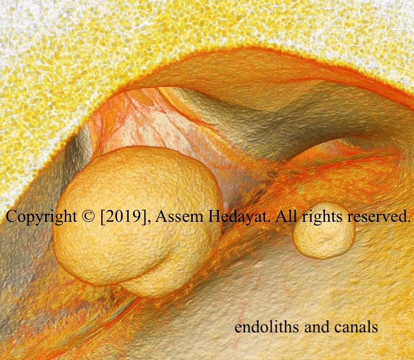
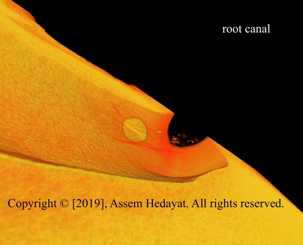
Volume rendering of pulp stones and root canal entrances
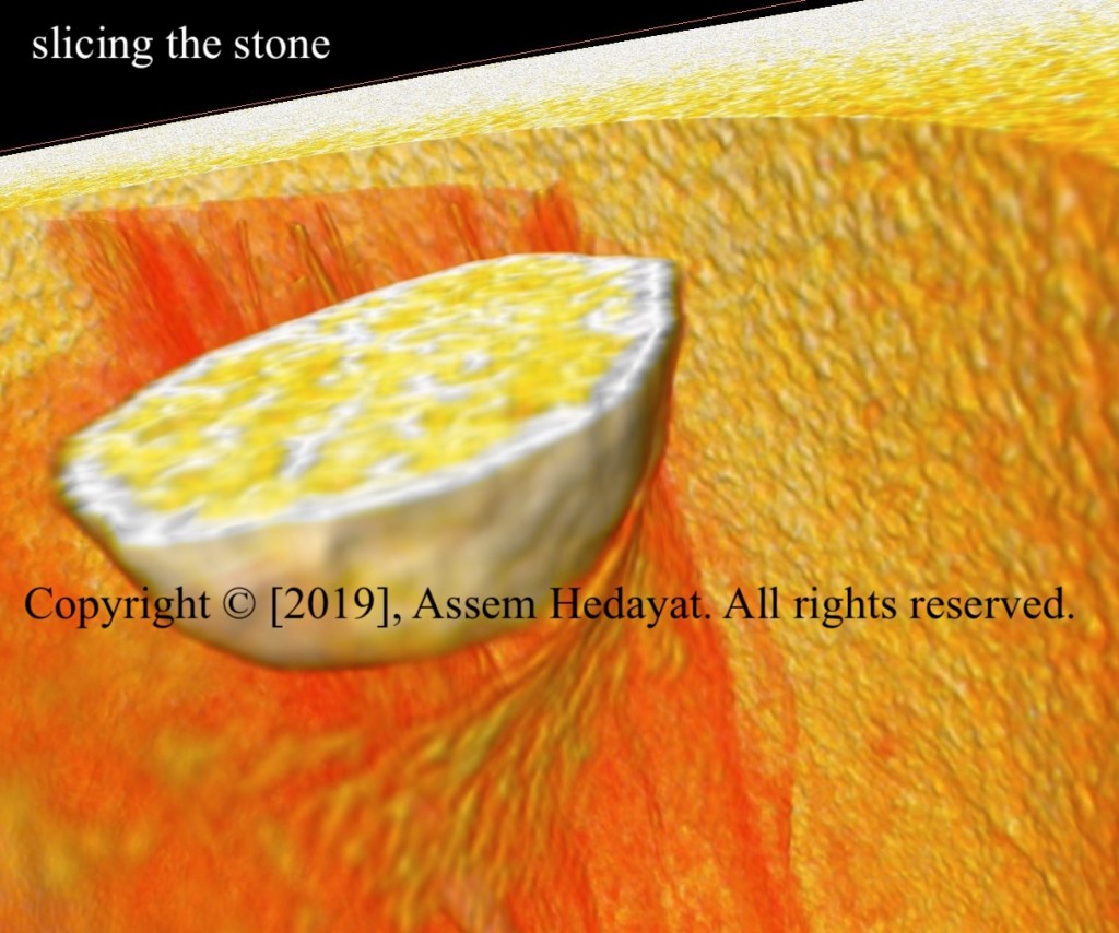
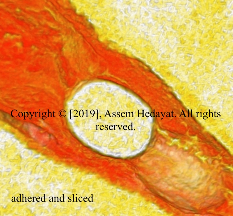
Volume rendering of a sliced root canal
About
Our team is led by Dr. Assem Hedayat, a highly accomplished biomaterials scientist with a Ph.D. from Michigan State University and a Master’s degree from the Georgia Institute of Technology.
Dr. Hedayat brings a wealth of academic and research expertise, having served as an Assistant Professor of Biomaterials at the College of Dentistry, University of Saskatchewan, Canada. His expertise lies in micro-CT imaging of teeth and dental restorative materials and their advanced 3D visualization—the very foundation of what we do.
Dr. Hedayat has shared his groundbreaking findings through peer-reviewed publications and has presented his research at national and international conferences, establishing himself as a trusted voice in the field of micro-CT imaging and volume rendering of dental structures and materials. He currently serves as an adjunct professor of Mechanical Engineering and is a member of the Division of Biomedical Engineering at the University of Saskatchewan, Canada.
Portfolio
A selection of our volume rendered images showcasing dental structures and restorative materials in stunning details.
For Dental Materials Companies:We partner with dental materials companies to support the improvement and development of their products through advanced 3D visualization. By rendering the internal structure of teeth alongside restorative materials, including composites, cements, and other dental materials, we provide manufacturers with an unprecedented view of how their products perform at the microscopic level. Our volume rendering capabilities deliver the precise visual insights needed to drive innovation, quality, and product excellence.
For the Dental Community: We proudly serve the broader dental community with stunning volume rendered images of dental structures and materials within for a wide range of professional needs, including conference and seminar presentations, annual reports and institutional statements, clinicians’ websites and digital profiles, printed materials such as brochures and posters, and research publications and journals.Whether you are a clinician, researcher, educator, publishing house or institution, our high-quality 3D dental imagery will elevate the visual impact of your professional materials.
For AI Dental Imaging and Diagnostics Companies & AI Platform Companies for Dental Histology: Our synchrotron images and 3D volume rendering of teeth provide a non-destructive, reproducible gold standard that no clinical imaging modality can replicate. They constitute benchmarking assets to validate algorithms against. This makes DentaDetail’s dataset uniquely valuable for AI that needs to learn true tooth anatomy. The 3D nature of the renders enables volumetric AI training that 2D histology slides and 2D radiographs simply cannot support. DentaDetail’s images can serve as the foundation for transfer learning. Starting with pre-training models on synchrotron data, clinical images will be fine tuned for real world deployment.